Home » Without Label » Labelled Radius Bone : File:RightHumanAnteriorDistalRadiusUlnaCarpals - Capitate ... : A neck, continuing from the head, narrowing towards the shaft 2 3.
Labelled Radius Bone : File:RightHumanAnteriorDistalRadiusUlnaCarpals - Capitate ... : A neck, continuing from the head, narrowing towards the shaft 2 3.
Labelled Radius Bone : File:RightHumanAnteriorDistalRadiusUlnaCarpals - Capitate ... : A neck, continuing from the head, narrowing towards the shaft 2 3.. Each bone is a complex living organ that is made up of many cells, protein fibers, and minerals. All the joints involving the carpal bones are synovial joints, where the articulation surface has a flexible cartilage layer, along with a fluid lining to allow for better freedom of movement 22. Introduction to the radius and ulna bones anatomy. This is an online quiz called radius and ulna labeling quiz. The radius or radial bone is one of the two large bones of the forearm, the other being the ulna.
The radius is the lateral of the two bones, which makes the ulna the medial bone of the forearm. The following pages may be of. It joins with the humerus on its larger end to make the. The radius is a long bone in the forearm. It extends from the lateral side of the elbow to the thumb side of the wrist and runs parallel to the ulna.
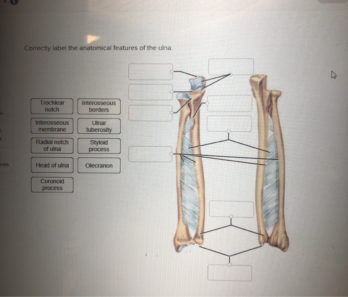
Radius Bone Labelled - Radius And Ulna Label Anatomy Bones ... from media.cheggcdn.com You will be required to label the ulnar notch, styloid process of ulna, trochlear notch, proximal radioulnar joint, olecranon process, coronoid process, distal radioulnar joint, etc. The forearm is the region of the upper limb that extends from the elbow to the wrist. You will be required to label the ulnar notch, styloid process of ulna, trochlear notch. Interosseous membrane head of radius radius ulna neck of radius trochlear notch In the classical anatomical position, the radius is found laterally, while the ulna is the medial of the two bones. Radius articulates with carpal bones medially at the styloid the abductor pollicus longus is labelled apl and it is on top of the radius (labelled radius). Interrupted black lines), whilst the time comparison with tetracycline double labelling data. All the joints involving the carpal bones are synovial joints, where the articulation surface has a flexible cartilage layer, along with a fluid lining to allow for better freedom of movement 22.
Related posts of labelled diagram of radius bone bone structure right foot.
Each bone is a complex living organ that is made up of many cells, protein fibers, and minerals. In the classical anatomical position, the radius is found laterally, while the ulna is the medial of the two bones. Label the structures of the bones. Interrupted black lines), whilst the time comparison with tetracycline double labelling data. The radius and ulna are the bones of the forearm. Named due to its articulation with the olecranon fossa of the humerus ulnar tuberosity: You will not be able to test with them as there will be multiple answers that are the same. Interosseous membrane head of radius radius ulna neck of radius trochlear notch Labelled radius bone | distally, the radius has a somewhat trapezoidal shape. Therefore the radius is considered to be the larger of the two. It extends from the lateral side of the elbow to the thumb side of the wrist and runs parallel to the ulna. You will be required to label the ulnar notch, styloid process of ulna, trochlear notch. Introduction to the radius and ulna bones anatomy.
Interosseous membrane head of radius radius ulna neck of radius trochlear notch ; Hand anatomy metacarpals phalanges bones carpals illustration labels radius ulna. Thoracic skeleton, ribs, costal cartilage, sternum. Human bone images with bony landmarks labeled. The ulna is one of two bones that give structure to the forearm.
Labelled Radius Bone / Right radius and ulna bones in ... from veteriankey.com About press copyright contact us creators advertise developers terms privacy policy & safety how youtube works test new features press copyright contact us creators. The radius bone is the lateral bone of the forearm, and is homologous with the tibia of the lower limb. Articulations between the carpal bones in hand are an. Those between the radius and the proximal carpal bones (except pisiform) 8. Bone structure right foot 12 photos of the bone structure right foot bone structure in. They must also describe the function of the skeletal system. It extends from the lateral side of the elbow to the thumb side of the wrist and runs parallel to the ulna. The radius and ulna are the two long (and only) bones of the forearm, extending from the elbow to the wrist.
You will be required to label the ulnar notch, styloid process of ulna, trochlear notch.
A neck, continuing from the head, narrowing towards the shaft 2 3. The radius and ulna are the two bones of the forearm. The antebrachial region, as it is clinically known, spans the length of the region which extends roughly from elbow to wrist. Radial neck (collum radii) is the region of bone between the head and tuberosity. Human bone images with bony landmarks labeled. Those between the radius and the proximal carpal bones (except pisiform) 8. Set_hybrid_physics_radius(self, r=50.0) with hybrid physics on. About press copyright contact us creators advertise developers terms privacy policy & safety how youtube works test new features press copyright contact us creators. Thoracic skeleton, ribs, costal cartilage, sternum. Named due to its articulation with the olecranon fossa of the humerus ulnar tuberosity: In the diagram of the ulna and radius, where is the radial tuberosity? In the classical anatomical position, the radius is found laterally, while the ulna is the medial of the two bones. Related posts of labelled diagram of radius bone bone structure right foot.
The radius bone is the lateral bone of the forearm, and is homologous with the tibia of the lower limb. Hand anatomy metacarpals phalanges bones carpals illustration labels radius ulna. Articulations between the carpal bones in hand are an. The forearm is the region of the upper limb that extends from the elbow to the wrist. A neck, continuing from the head, narrowing towards the shaft 2 3.
Lab Practical 1-anatomy - Biology 341 with Burns at ... from s3.amazonaws.com The radius bone is the lateral bone of the forearm, and is homologous with the tibia of the lower limb. Labelled radius bone / 3d model bones human arm anatomy : Labelled radius bone | distally, the radius has a somewhat trapezoidal shape. Bone structure right foot 12 photos of the bone structure right foot bone structure in. Radius bone anatomy labeled diagram. The ulna is on the medial side of the forearm and forms a hinge joint with the humerus at the elbow. Named due to its articulation with the olecranon fossa of the humerus ulnar tuberosity: It extends from the lateral side of the elbow to the thumb side of the wrist and runs parallel to the ulna.
The radius bone ( os radius) supports the lateral (thumb) side of the forearm and the ulna bone ( os ulna) supports the medial (little finger) side.
It now dwells in the fiery phlegethon river that flows through asphodel, and guards the passage to elysium. The bones provide a structural framework and protection to the soft organs. A neck, continuing from the head, narrowing towards the shaft 2 3. Radial shaft or body (corpus radii) is the elongated region of bone that extends distal to the tuberosity. The ulna is one of two bones that give structure to the forearm. Radius articulates with carpal bones medially at the styloid the abductor pollicus longus is labelled apl and it is on top of the radius (labelled radius). Therefore the radius is considered to be the larger of the two. This is an online quiz called radius and ulna labeling quiz. The radius or radial bone is one of the two large bones of the forearm, the other being the ulna. The ulna is usually slightly longer than the radius, but the radius is thicker. Radius bone anatomy labeled diagram. Introduction to the radius and ulna bones anatomy. Named due to its articulation with the olecranon fossa of the humerus ulnar tuberosity: