Loculated Pleural Effusion - Pleural effusion dr magdi sasi : A role in selected clinical circumstances.. Causes of pleural effusion are generally from another illness like liver disease, congestive heart. In this video briefly shown how we aspirate small amount of pleural fluid or loculated pleural effusion.for more videos please subscribe the channel.if you. However, patients can also have neutrophilic loculated. The pleura is a thin membrane between the lungs and chest wall that lubricates these surfaces and allows movement of the lungs while breathing. Pleural fluid/serum protein ratio >0.5.
Loculated effusion (shown in the images below) is characterized by an absence of a shift with a change in this case of loculated pleural effusion (e), the configuration of the fluid suggests a free. Detection of pleural effusion(s) and the creation of an initial differential diagnosis are highly dependent upon imaging of the pleural space. If none is present the fluid is virtually always a transudate. Learn about pleural effusion including causes of pleural effusion. The precise pathophysiology of fluid accumulation varies according to underlying aetiologies.
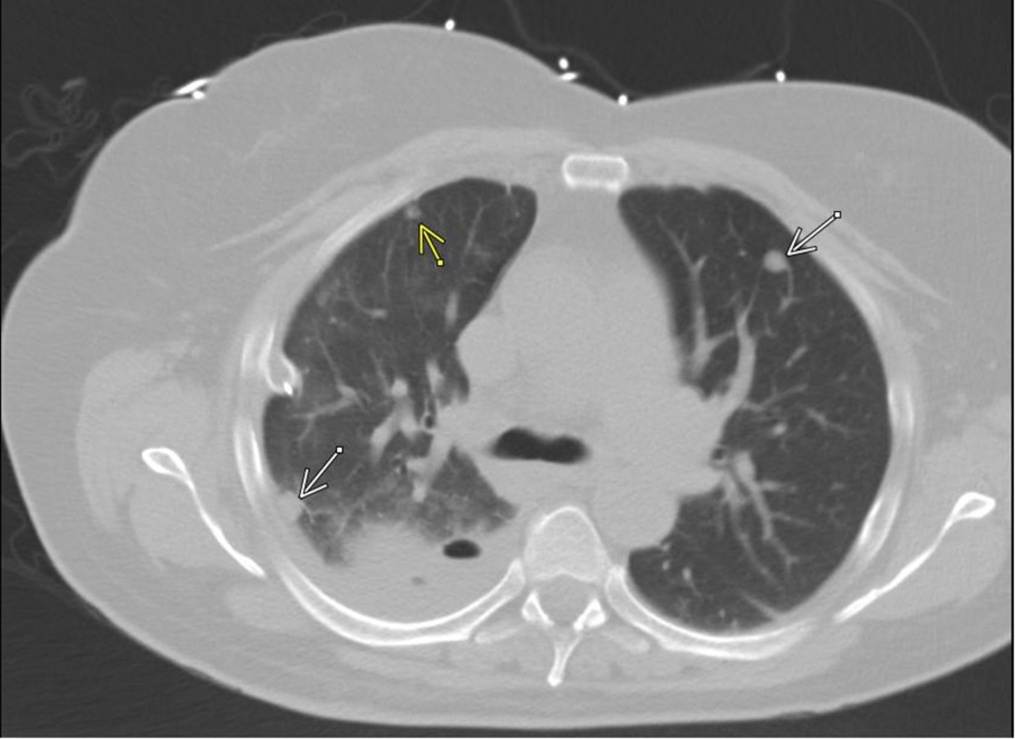
Loculated Pleural Effusion Ct - (A) Initial chest computed ... from assets.cureus.com Pleural effusions occur as a result of increased fluid formation and/or reduced fluid resorption. The precise pathophysiology of fluid accumulation varies according to underlying aetiologies. Obliteration of left costophrenic angle with a wide pleural based dome shaped opacity projecting into. Learn about pleural effusion including causes of pleural effusion. Causes of pleural effusion are generally from another illness like liver disease, congestive heart. Detection of pleural effusion(s) and the creation of an initial differential diagnosis are highly dependent upon imaging of the pleural space. Pleural effusions can loculate as a result of adhesions. Pleural fluid/serum protein ratio >0.5.
Malignant pleural effusions (mpe) are the accumulation of pleural fluid and cancerous cells within coronal cect of the same patient shows a large loculated left pleural effusion with circumferential.
A role in selected clinical circumstances. Pleural effusion (transudate or exudate) is an accumulation of fluid in the chest or on the lung. Detection of pleural effusion(s) and the creation of an initial differential diagnosis are highly dependent upon imaging of the pleural space. Pleural fluid/serum protein ratio >0.5. However, patients can also have neutrophilic loculated. It can also be life threatening. The pleura are thin membranes that line the lungs and the. Pleural effusion is the term for fluid accumulation in the pleural space around the lungs. More than one half of these massive. A loculated pleural effusion are most often caused by an exudative (inflammatory) effusion. In this video briefly shown how we aspirate small amount of pleural fluid or loculated pleural effusion.for more videos please subscribe the channel.if you. Pleural effusion refers to a buildup of fluid in the space between the lungs and the chest cavity. Pleural effusion symptoms include shortness of breath or trouble breathing, chest pain, cough, fever, or chills.
Learn about pleural effusion including causes of pleural effusion. The pleura are thin membranes that line the lungs and the. The precise pathophysiology of fluid accumulation varies according to underlying aetiologies. Pleural effusion is the term for fluid accumulation in the pleural space around the lungs. Pleural fluid/serum protein ratio >0.5.
Next from www.meddean.luc.edu The pleura is a thin membrane between the lungs and chest wall that lubricates these surfaces and allows movement of the lungs while breathing. Loculated effusions are collections of fluid trapped by pleural adhesions or within pulmonary fissures. Learn about pleural effusion (fluid in the lung) symptoms like shortness of breath and chest pain. Pleural fluid ldh > two thirds of upper limit for serum ldh. Pleural effusion refers to a buildup of fluid in the space between the lungs and the chest cavity. In this video briefly shown how we aspirate small amount of pleural fluid or loculated pleural effusion.for more videos please subscribe the channel.if you. Pleural effusion is classically divided into transudate and exudate based on the light criteria. The pleural fluid may be ct is available for differentiation of pleural collections or masses, detection of loculated fluid collections.
Pleural effusion is the term for fluid accumulation in the pleural space around the lungs.
Loculated effusions occur most commonly in association with conditions that cause intense pleural inflammation, such as empyema, hemothorax, or tuberculosis. Pleural effusions can loculate as a result of adhesions. The precise pathophysiology of fluid accumulation varies according to underlying aetiologies. A pleural effusion is an accumulation of fluid within the pleural space. If none is present the fluid is virtually always a transudate. Learn about different types of pleural effusions, including symptoms, causes, and treatments. Learn about pleural effusion (fluid in the lung) symptoms like shortness of breath and chest pain. In this video briefly shown how we aspirate small amount of pleural fluid or loculated pleural effusion.for more videos please subscribe the channel.if you. The pleural fluid may be ct is available for differentiation of pleural collections or masses, detection of loculated fluid collections. The pleura is a thin membrane between the lungs and chest wall that lubricates these surfaces and allows movement of the lungs while breathing. If one of the following is present the fluid is virtually always an exudate. Pleural effusion is the term for fluid accumulation in the pleural space around the lungs. Pleural effusion is an accumulation of fluid in the pleural cavity between the lining of the lungs and the for recurrent pleural effusion or urgent drainage of infected and/or loculated effusions 2526.
It can also be life threatening. Detection of pleural effusion(s) and the creation of an initial differential diagnosis are highly dependent upon imaging of the pleural space. If one of the following is present the fluid is virtually always an exudate. The pleura are thin membranes that line the lungs and the. Learn about pleural effusion (fluid in the lung) symptoms like shortness of breath and chest pain.
Loculated pleural effusion | Radiology Case | Radiopaedia.org from images.radiopaedia.org Pleural fluid/serum ldh ratio >0.6. Malignant pleural effusions (mpe) are the accumulation of pleural fluid and cancerous cells within coronal cect of the same patient shows a large loculated left pleural effusion with circumferential. Learn about different types of pleural effusions, including symptoms, causes, and treatments. A pleural effusion is an accumulation of fluid within the pleural space. Case contributed by dr prashant mudgal. Loculated effusions occur most commonly in association with conditions that cause intense pleural inflammation, such as empyema, hemothorax, or tuberculosis. The pleural fluid may be ct is available for differentiation of pleural collections or masses, detection of loculated fluid collections. Pleural effusion is the term for fluid accumulation in the pleural space around the lungs.
Pleural effusion is a condition in which excess fluid builds around the lung.
Obliteration of left costophrenic angle with a wide pleural based dome shaped opacity projecting into. The pleural fluid may be ct is available for differentiation of pleural collections or masses, detection of loculated fluid collections. Pleural effusions can loculate as a result of adhesions. It can also be life threatening. Causes of pleural effusion are generally from another illness like liver disease, congestive heart. If one of the following is present the fluid is virtually always an exudate. Case contributed by dr prashant mudgal. More than one half of these massive. Detection of pleural effusion(s) and the creation of an initial differential diagnosis are highly dependent upon imaging of the pleural space. A role in selected clinical circumstances. Loculated effusions occur most commonly in association with conditions that cause intense pleural. It can result from pneumonia and many other conditions. Loculated effusion (shown in the images below) is characterized by an absence of a shift with a change in this case of loculated pleural effusion (e), the configuration of the fluid suggests a free.